Canadian Viagra
Men's Blog from Canada
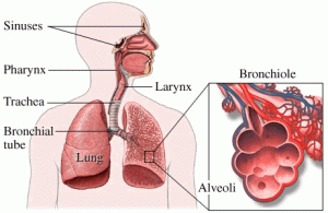
Lung function in patients with asthma
19 Jan 2015There are several studies showing the relationship between the phenotypes of airway inflammation and remodeling. It has been reported that eosinophilic asthma has more RBM thickening and airflow obstruction than noneosinophilic asthma. Higher neutrophil counts in sputum correlate with airflow obstruction. Airway eosinophilia and neutrophilia are independent of each other in asthma.
Lung function
Although causal relationships are hard to establish, the frequency and reproducibility of association with lung function argue strongly for an influence on lung function. The increase of smooth muscle mass, a consistent feature of airway remodeling, correlates with lower lung function in patients with asthma. In COPD, the more extensive the pathologic changes, including increase of ASM, squamous cell and goblet cell metaplasia, and fibrosis, the lower is lung function. Patients with asthma and COPD with fixed airflow obstruction exhibited a more rapid decline in lung function and experienced more frequent exacerbations in a 5-year follow-up compared with patients with asthma with reversible airflow obstruction. In cystic fibrosis, the increase of inner wall and smooth muscle area are even greater than COPD, and these dimensional changes presumably contribute to severe airflow limitation.
Some investigators have argued for a favorable effect of some aspects of remodeling. For example, airway wall stiffness due to RBM thickening was reported to reduce mucosal folding during smooth muscle contraction, diminishing the magnitude of airway obstruction. The advantages to the host of many aspects of airway remodeling are hard to argue.
The improvement of the resolution by CT scanning has enabled the measurement of airway dimensions in relatively large airways. Measuring airway wall thickness remains a challenge, and because CT scanning is incapable of analyzing the airway composition, it cannot distinguish between true increase in airway wall thickness from remodeling and the effects of bronchoconstriction that will increase airway area for a given lumen size. There are emerging techniques that have been applied during bronchoscopy to assess airway structure. Optical coherence tomography, a light-based imaging technique, has been used to quantify large airways distensibility, and fibered confocal fluorescence microscopy has been used to visualize elastic fibers in central airway walls.
About this blog
Lorem ipsum dolor sit amet, consectetuer adipiscing elit. Quisque sed felis. Aliquam sit amet felis. Mauris semper, velit semper laoreet dictum, quam diam dictum urna, nec placerat elit nisl in quam. Etiam augue pede, molestie eget, rhoncus at, convallis ut, eros. Aliquam pharetra.
Photostream
Categories
- Asthma (1)
- Canadian Health Care (1)
- Canadian Viagra (1)
- Disorders (1)
- Erectile Dysfunction (1)
- Health and Care (2)
- HQ Canadian Pharmacy (1)
- Medicine (1)
- No comments
- Viagra Online at Canadian Pharmacy
- How to Cure Erectile Dysfunction in Male with Canadian Viagra?
- Lung function in patients with asthma
- Patients treated with high-dose ICSs
- HQ Canadian Pharmacy – Imagination
- Disorders Identification Test Consumption
- Risks and Rewards of Responsibility
- Nourishing the Soil
Comments are closed.